
Differences among X-rays, CT, B-mode ultrasound
 May 14, 2016|
 View:473
X -rays ---- radiograph
Bones contain much calcium, which due to its relatively high atomic numberabsorbs x-rays efficiently. This reduces the amount of X-rays reaching the detector in the shadow of the bones, making them clearly visible on the radiograph. The lungs and trapped gas also show up clearly because of lower absorption compared to tissue, while differences between tissue types are harder to see.
Advantages: convenient and cheap
Disadvantages: it has superimposition of images of tissues so it will be seen clearly when being shot for many times and from different angles.
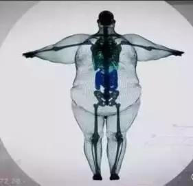
( A man with 900kg under X-rays )
C T
It makes use of computer-processed combinations of many X-ray images taken from different angles to produce cross-sectional (tomographic) images (virtual "slices") of specific areas of a scanned object, allowing the user to see inside the object without cutting.
Advantages: it is tomographic so we can get more information after disposal
Disadvantages: it is more expensive than X-rays and the radiation doses received from CT scans are 100 to 1,000 times higher than conventional X-rays
( 3D reconstruction image of a chopstick inserted to the eye )
B-mode ultrasound
Ultrasonic images also known as sonograms are made by sending pulses of ultrasound into tissue with a probe.
Advantages: polydirectional observation and real time imagery
Disadvantages: it is easy to be disturbed by gas and the accuracy rate will decrease when observe organs with many gas, such as intestinal canal and so on. So, enteroscope is often used to observe intestinal canal
Magnetic Resonance Imaging ( MRI )
It uses the signals through collecting MRI to build up enough information to construct an image of body. That is to say, shake and vibrate hydrogen protons and then feel the vibration.
Advantages: Compared to CT, it does not have radioactive radiation and bony artifact. It will be imaging in multiparameter and many ways. It also has altitudinal ability to distinguish soft tissues.
Disadvantages: expensive

( carambola under MRI )
Different ways for different parts of body
1、Traumatic Bones——a rough look in X-rays and a scan in CT. Choose X-rays first because it is convenient and easy to get results. If you want more inspection, you can choose CT ,or MRI to examine hidden hurt or soft tissues hurt.
2、Cervical and Lumber Vertebra——2.First MRI and then CT. Cervical spondylosis and prolapse of lumbar intervertebral disc ( PLID ) need to examine intervertable disc and relevant nerve root. If you want to examine these soft tissues better, the best choice is MRI. Similarly, the examination of joints, muscles, adipose tissues, tumour, inflammation, trauma, degenerative changes and various congenital diseases, MRI is also a good choice. The examination of vertebral bone hyperplasia and intervertebral foramina stenosis can also use CT.
3、Chest——A rough look in X-rays and a scan in CT. X-ray chest image can have a rough look in lungs, podoid, arcus aorta, ribs and so on. It can also examine whether lung markings increase, big lumps in lung, aortic calcification and so on. CT chest image shows clearer structure. It is more accurate and sensitive than X-rays when finding out chest lesions, especially meaningful to screening early stage of lung cancer. MRI is limited to diagnose lung diseases.
4、Abdomen and Pelvic Cavity——B-mode ultrasound can examine all the organs except intestinal canals. As we all known, the examination of fetus always uses B-mode ultrasound. Besides, B-mode ultrasound has high accuracy to examine superficial organs, such as thyroid, and parenchymatous organs, such as livers, spleens, pancreas, kidneys and pelvic cavities.
5、Heart——Unless CT is applied to coronary heart disease, and B-mode ultrasound is applied to cardiac function, common examination of heart structure and function often uses cardiac color ultrasound. CT can also be used to examine coronary artery and congenital heart disease in structural abnormality. MRI is used to examine myocardial lesions, such as myocardial infarction.
 WhatsApp:
 E-mail:
 Mobile: